
Chromosomes are the essential structures that store and transmit genetic information in all living organisms. In humans, these microscopic entities play a crucial role in determining our traits, health, and overall development. A photograph of an individual’s chromosomes can reveal intricate details about their genetic makeup, which can have profound implications in the fields of genetics, medicine, and anthropology. In this article, we will delve into the world of chromosomes, exploring their structure, function, and significance, and how these can be visualized through techniques like karyotyping.
Exploring the captivating realm of chromosomal imaging, this remarkable endeavor unveils the intricate world of genetics and cell biology. Precision and advanced imaging techniques unlock the mysteries hidden within our cells, offering a glimpse into the very essence of human life at a molecular level. Join us on this scientific journey to understand how to capture photographs of individual chromosomes? and the technology that powers this discovery.
A photograph of an individual’s chromosomes captures their genetic blueprint. These microscopic structures, often seen as X-shaped, carry essential information. Karyotyping is the technique used to visualize and analyze chromosomes. It’s vital for diagnosing genetic disorders and understanding genetic diversity. This glimpse into our genetic makeup holds profound significance in the fields of medicine and genetics.
The Chromosomal Basis of Life
The Chromosomal Basis of Life refers to the fundamental role that chromosomes play in the storage and transmission of genetic information, shaping the characteristics and traits of all living organisms. Understanding this genetic foundation is pivotal in the field of genetics and biology.
Chromosomes as Genetic Blueprints
Chromosomes are thread-like structures found within the nucleus of eukaryotic cells, where they carry the genetic information necessary for the development, growth, and functioning of an organism. In humans, the genetic blueprint is stored in 46 chromosomes, divided into 23 pairs, one from each parent.
Human Chromosome Pairs
Of these 23 pairs, 22 pairs are autosomes, and one pair consists of sex chromosomes, determining an individual’s gender. The 22 autosomal pairs are virtually identical in both males and females, but the sex chromosomes differ significantly. Males have one X and one Y chromosome (XY), while females have two X chromosomes (XX).
The Structure of Chromosomes
The structure of chromosomes in a cell is comprised of long, coiled strands of DNA, which are intricately organized with proteins. This compact and organized structure capture a photograph of an individual’s chromosomes is essential for the storage and accurate transmission of genetic information during cell division.
DNA, The Building Block of Chromosomes
Chromosomes are primarily composed of deoxyribonucleic acid (DNA), which is a long, double-stranded molecule. DNA is organized into a compact structure, which we commonly refer to as chromatin. Chromatin consists of DNA wrapped around histone proteins, forming nucleosomes.
The X-Shaped Chromosome
When viewed under a microscope, chromosomes often appear as X-shaped structures, especially during cell division. This distinctive shape is due to the coiling and condensation of the chromatin, which ensures the genetic material can be accurately duplicated and distributed during cell division.
Visualizing Chromosomes, Karyotyping
Visualizing chromosomes through karyotyping is a powerful technique in genetics and cytogenetics, enabling scientists to visually inspect and analyze an individual’s complete set of chromosomes, providing valuable insights into genetic abnormalities and variations.
Karyotyping, A Window into Genetic Information
Karyotyping is a laboratory technique used to visualize and analyze a photograph of an individual’s chromosomes . It involves photographing the chromosomes when they are most condensed, typically during cell division. This technique is invaluable in diagnosing genetic disorders, understanding genetic diversity, and even solving crimes in forensics.
The Karyotyping Process
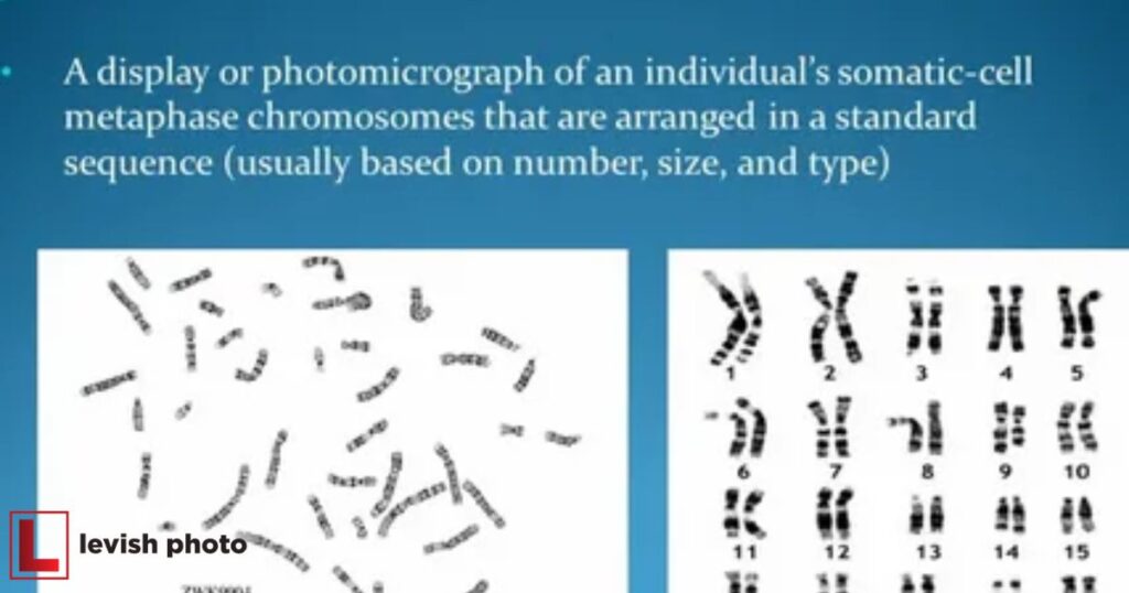
The karyotyping process begins with a sample of an individual’s cells, often extracted from blood, amniotic fluid, or bone marrow. These cells are then cultured and treated with chemicals to arrest them in the metaphase stage of cell division when chromosomes are most visible. After this, the cells are stained to create distinct bands on the chromosomes, making them easier to differentiate and identify.
Interpreting a Karyotype
Interpreting a karyotype involves analyzing an individual’s chromosome arrangement, size, and number to identify genetic abnormalities, such as aneuploidies or structural variations. This visual representation of an individual’s genome is a crucial tool in diagnosing genetic disorders and understanding chromosomal health.
The Karyotype Image
A karyotype is a photographic representation of an individual’s chromosomes arranged in pairs according to their size, shape, and banding patterns. The pairs are ordered from the largest (chromosome 1) to the smallest (chromosome 22), with the sex chromosomes (X and Y) being the last pair.
Trisomy and Monosomy
Karyotyping can reveal chromosomal abnormalities, such as trisomy, where an individual has an extra chromosome in a pair (e.g., Down syndrome with three copies of chromosome 21), or monosomy, where a chromosome is missing (e.g., Turner syndrome with only one X chromosome). These abnormalities can lead to developmental and health issues.
Sex Chromosome Disorders
Issues with sex chromosomes are also easily identified through karyotyping. Conditions like Klinefelter syndrome (XXY) and Turner syndrome (X) result from sex chromosome abnormalities, affecting sexual development and overall health.
Genetic Disorders and Karyotyping
Genetic disorders, often caused by chromosomal abnormalities, can have a profound impact on an individual’s health. Karyotyping, a diagnostic technique that analyzes an individual’s chromosomes, plays a crucial role in identifying such genetic anomalies, aiding in diagnosis and treatment decisions.
Down Syndrome
Down syndrome, also known as Trisomy 21, is one of the most well-known chromosomal disorders. It is caused by an extra copy of chromosome 21 and is often associated with intellectual disabilities, distinctive physical features, and various health problems.
Turner Syndrome
Turner syndrome, on the other hand, is a condition where one of the X chromosomes is missing in females. It results in short stature, infertility, and various health issues. Karyotyping is an essential tool for diagnosing Turner syndrome and determining the genetic basis for these characteristics.
Klinefelter Syndrome
Klinefelter syndrome is characterized by the presence of one or more extra X chromosomes in males (e.g., XXY). This condition can lead to infertility, hormonal imbalances, and developmental issues. Karyotyping is instrumental in identifying the chromosomal abnormalities associated with Klinefelter syndrome.
Karyotyping Beyond Genetic Disorders
Karyotyping extends beyond the realm of genetic disorders, finding a crucial role in prenatal diagnostics. By identifying chromosomal abnormalities in developing fetuses, it empowers parents and healthcare professionals to make informed choices regarding pregnancy and medical interventions.
Prenatal Diagnostics
Karyotyping is a valuable tool in prenatal diagnostics. It can detect chromosomal abnormalities in a developing fetus, allowing parents and healthcare providers to make informed decisions about pregnancy, medical care, and potential interventions.
Forensic Applications
Karyotyping is also used in forensic science to solve criminal cases. By analyzing the chromosomes present in evidence such as hair, blood, or semen, forensic experts can identify suspects and victims.
| Aspect | Description |
| Karyotype Image | Photographic representation of individual chrommes in pairs, ordered by size, shape, and banding patterns, with sex chromosomes (X and Y) being the last pair.oso |
| Trisomy and Monosomy | Karyotyping identifies chromosomal abnormalities: trisomy (extra chromosome) and monosomy (missing chromosome), affecting development and health. |
| Sex Chromosome Disorders | Detects issues with sex chromosomes, like Klinefelter syndrome (XXY) and Turner syndrome (X), impacting sexual development and overall health. |
| Genetic Disorders and Karyotyping | Essential in diagnosing conditions such as Down Syndrome (Trisomy 21), Turner Syndrome, and Klinefelter Syndrome, each with unique characteristics. |
| Karyotyping Beyond Genetic Disorders | Valuable in prenatal diagnostics, allowing early detection of chromosomal abnormalities in fetuses. Used in forensic science to identify suspects and victims by analyzing chromosomes in evidence like hair, blood, or semen. |
The Future of Chromosome Research
Advances in Genetic Testing
As technology continues to advance, genetic testing methods are becoming more precise and accessible. Techniques like next-generation sequencing (NGS) are supplementing and, in some cases, replacing traditional karyotyping, allowing for faster and more comprehensive analysis of an individual’s genetic material.
Personalized Medicine
Understanding an individual’s chromosomal makeup is crucial in the field of personalized medicine. It can guide the development of tailored treatments and therapies, taking into account a person’s unique genetic characteristics.
Ethical Considerations
The increasing availability of genetic information through techniques like karyotyping raises ethical questions about privacy, discrimination, and the potential for misuse of genetic data. These issues will continue to be important topics of discussion as technology advances.
FAQs
What are chromosomes and why are they important?
Chromosomes store genetic information and are crucial for determining an individual’s traits and health.
What is karyotyping, and how is it used in genetics?
Karyotyping is a technique that visualizes an individual’s chromosomes, aiding in diagnosing genetic disorders and understanding diversity.
How is karyotyping impacting fields beyond genetics?
Karyotyping is employed in prenatal diagnostics, forensics, and personalized medicine, making it a versatile tool in various domains.
Conclusion
In conclusion, the ability to capture a photograph of an individual’s chromosomes, achieved through the technique of karyotyping, unveils a profound window into the world of genetics. This revolutionary method has not only deepened our comprehension of human biology but also played a pivotal role in the diagnosis of genetic disorders, the advancement of prenatal diagnostics, and the support of forensic investigations.
As technology continues to progress, the domains of medicine, genetics, and anthropology are on the brink of unprecedented breakthroughs. Armed with these tools, we stand poised to further explore and decipher the intricate genetic tapestry that makes each of us a unique and complex individual.