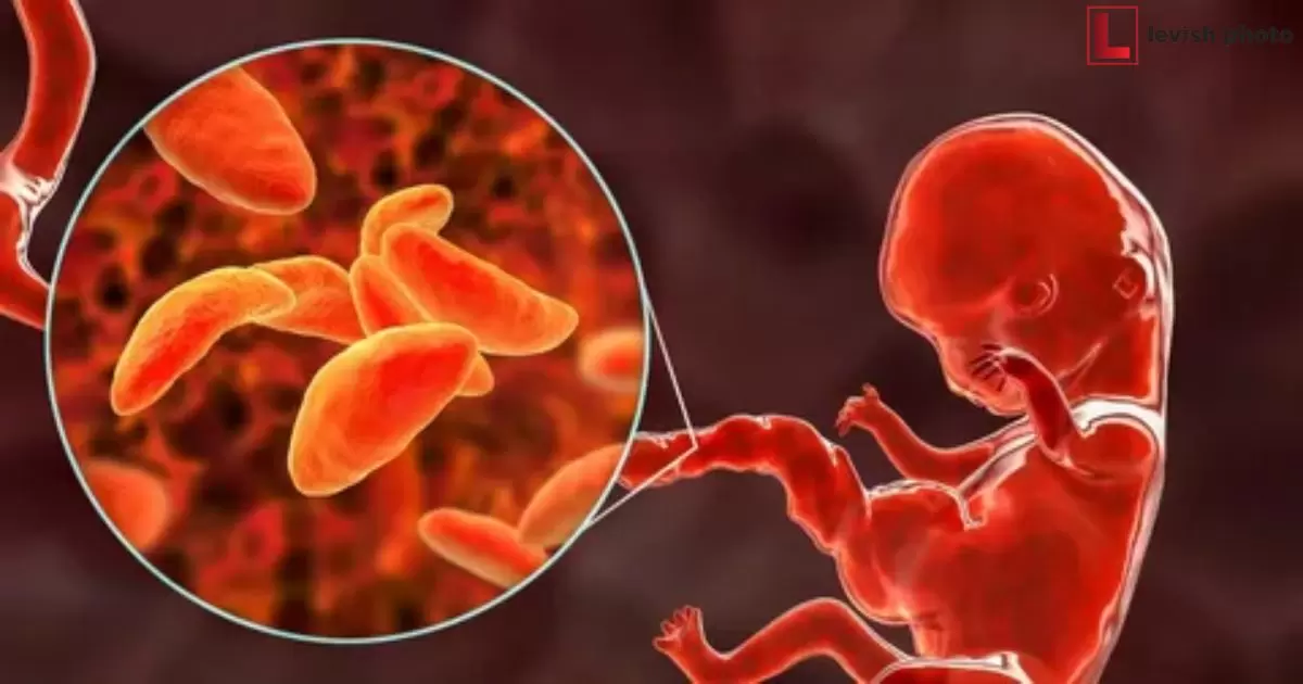
An atlas of developmental biology, in the context of our topic, is a visual reference tool that showcases the various stages and processes of embryonic development. It’s like a map, but instead of geography, it guides us through the intricate journey of life’s creation, providing a unique insight into the growth and transformation of organisms.
Imagine flipping through the pages of a photographic atlas of developmental biology, each image capturing the magic of life unfolding. It’s a captivating journey through the early stages of existence, a visual adventure that brings the wonders of biology to life right before your eyes.
In this fascinating atlas of developmental biology, you’ll find a collection of meticulously documented images that unveil the mysteries of how organisms form and mature. These snapshots of life’s earliest moments offer invaluable knowledge for scientists, students, and anyone curious about the miracle of existence.
Historical Perspective
We delve into the evolution of developmental biology imaging techniques and the development of photographic atlases over the years. This segment explores the early methods and technologies used for visualizing the intricate processes of embryonic development, tracing back to the rudimentary microscopy and illustration techniques employed by early biologists.
This historical context helps readers understand the transformation in the field and the contributions of early pioneers in developmental biology imaging, setting the stage for the discussion of modern photographic atlases and their importance in contemporary research and education.
Creating a Photographic Atlas
Creating a Photographic Atlas is a pivotal aspect of the field of developmental biology, serving as a visual repository of meticulously captured images that document various stages of organism development. This section of the article delves into the intricate process of assembling such atlases, beginning with the critical step of Sample Preparation.
Where specimens are carefully collected, preserved, and sometimes stained to highlight specific features. The subsequent segment explores the choice of Imaging Techniques and Equipment, emphasizing the need for cutting-edge microscopes, cameras, and imaging software to capture high-resolution, accurate images.
Data Processing and Analysis are discussed, highlighting the critical role of image processing to enhance clarity and extract quantitative data from the raw images. Together, these elements ensure that a photographic atlas is not only a visual masterpiece but also a valuable scientific resource for researchers, educators, and students in the field of developmental biology.
Key Stages of Development
| Developmental Stage | Description |
| Fertilization | The initial stage where a sperm cell fertilizes an egg, resulting in the formation of a zygote. |
| Cleavage | Involves a series of rapid cell divisions, leading to the formation of a multicellular embryo. |
| Gastrulation | The process during which the germ layers (ectoderm, mesoderm, and endoderm) are established. |
| Organogenesis | The stage where the organs and tissues of the organism begin to develop and take shape. |
| Tissue Differentiation and Growth | Cells become specialized, and the organism matures, growing and differentiating into specific cell types and structures. |
These stages represent the fundamental events that occur during the development of organisms and provide critical insights into the processes underlying life’s beginnings.
Model Organisms
Model Organisms refers to specific species that are commonly used in scientific research to investigate various aspects of biology, including developmental biology. These organisms are chosen for their well-defined genetics, short generation times, and amenability to laboratory manipulation.
In the context of a photographic atlas of developmental biology, model organisms such as zebrafish, Drosophila (fruit flies), Xenopus (African clawed frogs), mice, and human embryos play a crucial role. They offer insights into the conserved processes of embryonic development and allow scientists to explore the molecular and cellular mechanisms that underlie key developmental stages.
By using these model organisms, researchers can create detailed visual records of embryonic development, helping to uncover the intricacies of how organisms develop from a single cell into a complex, multicellular organism.
Applications and Significance
Applications and Significance explores the multifaceted impact of photographic atlases in developmental biology. These atlases, a photographing tool used for arguments, play a crucial role in education and research, serving as invaluable resources for students and scientists alike by providing visual insights into the intricate processes of embryonic development.
Beyond academia, they have substantial clinical and medical applications, aiding in the understanding of birth defects, developmental disorders, and regenerative medicine. Photographic atlases provide evolutionary insights, helping researchers trace the origins of various biological processes.
Their visual documentation of developmental stages across different species offers a comparative perspective, shedding light on the conserved and divergent aspects of embryogenesis. These atlases serve as a bridge between fundamental science and practical applications, making them indispensable tools for advancing our knowledge of life’s beginnings.
Notable Photographic Atlases
Notable Photographic Atlases provides an overview of some of the most influential and noteworthy publications in the field of developmental biology imaging. This section delves into landmark works that have significantly contributed to our understanding of embryonic development and tissue differentiation, showcasing their pivotal role in shaping the field.
It may include discussions on iconic atlases such as The Atlas of Human Development, The Zebrafish Book, and other seminal publications, highlighting their impact on both education and research in developmental biology. This section is a recognition of the valuable resources that have been instrumental in advancing the knowledge of development over the years.
Challenges and Future Directions
As the field of developmental biology continues to advance, it faces several challenges and opportunities for future growth. These challenges encompass technological, ethical, and legal aspects of research and imaging.
| Challenges | Future Directions |
| Technological Advancements | Development and integration of more advanced imaging techniques, such as super-resolution microscopy and 3D tissue reconstruction, to capture finer details of embryonic development. |
| Data Management | Addressing the increasing volume of data generated by high-resolution imaging, necessitating the development of robust data storage and analysis methods. |
| Ethical and Legal Considerations | Developing guidelines and regulations for the use of human embryos in research, as well as addressing ethical concerns related to genetic manipulation and embryo development. |
| Accessibility and Education | Ensuring that photographic atlases are widely accessible to researchers, educators, and students worldwide, promoting a broader understanding of developmental biology. |
| Interdisciplinary Collaboration | Encouraging collaboration between biologists, computer scientists, and experts from other fields to harness the full potential of technology and data analysis in developmental biology. |
| Long-term Studies | Supporting research that spans extended periods to capture the dynamic nature of development, potentially through improved embryo culture techniques. |
| Public Engagement | Enhancing public awareness and understanding of developmental biology and the ethical considerations surrounding it through outreach and education. |
These challenges, when addressed, have the potential to open new frontiers in our understanding of developmental biology and its applications in fields such as regenerative medicine and evolutionary biology.
Frequently Asked Questions
What are the primary challenges in developing photographic atlases of developmental biology?
Challenges include technological limitations, ethical considerations, data management, and the need for interdisciplinary collaboration.
How do technological advancements impact the future of developmental biology imaging?
Technological advancements enable the capture of finer details in embryonic development, fostering a deeper understanding of the subject.
What role do model organisms play in developmental biology photographic atlases?
Model organisms serve as valuable subjects for studying developmental processes and provide insights applicable to humans and other species.
Why is it important to address ethical and legal considerations in developmental biology research?
Addressing these considerations ensures responsible and ethical research practices while navigating the complexities of embryo development.
How can the public engage with and learn from developmental biology photographic atlases?
Public engagement can be fostered through outreach and education to increase awareness and understanding of developmental biology and its ethical dimensions.
Conclusion
In the world of developmental biology, photographic atlases serve as crucial tools to unlock the secrets of life’s beginnings. They capture the intricate dance of cells, tissues, and organs as they form and grow. Through these visual records, scientists gain deeper insights into the complexities of development, offering potential breakthroughs in regenerative medicine, understanding evolution, and more.
As we navigate the challenges and embrace the promise of technological advancements, ethical considerations, and broader accessibility, the future of developmental biology imaging shines brightly. The collaborative efforts of biologists, data scientists, and educators will continue to propel this field forward, ensuring that the beauty and mystery of life’s creation are shared and understood by all.